Microscopy
Elemental analysis of a fluid sample by Inductively Coupled Plasma Spectrometer (ICP) is limited to very small particles, usually less than 5μm (microns) in size; microscopic analysis is suitable for identifying particles from 3μm to over 1,000μm.
Microscopy is the only practical method of determining the wear mechanism which produced the debris found in a sample.
Filtergrams
Filtergrams are the most comprehensive test our laboratory offers for analysing particulate wear debris.
The sample to be examined is dissolved in a solvent and filtered through a very fine membrane filter. The wear debris and other contaminants in the oil are captured on the membrane for examination under a high power microscope. The filtergram report gives an analysis of the size and composition of all the particles found as well as the wear mechanism which generated the metallic particles.
Photographs of selected particles are presented on the report. Debris recovered from filter media can also be analysed by these techniques.
Rapid Filtergrams
Rapid Filtergrams involve analysis of the sample under a Microscope which also includes a quantification of the material, wear mechanism and size. This test will also include a representative image of the sample, as taken under the microscope, typically at 500x magnification. A graph also accompanies the image, plotting the wear mechanism against the size, showing the overall severity of the contamination.
Filter Patches
The filter patch is a quick method of extracting the wear debris for microscopic examination and comparison with other routine test results performed on the sample.
For example, this test can be used to confirm the presence of copper leaching as opposed to mechanical wear of a copper alloy component.
The SEM offers the potential to positively identify the alloy composition of individual wear particles.
Ferrograms
The ferrogram is a technique to prepare samples for microscopic examination by separating ferrous wear particles in a magnetic field. This will sort the particles according to their size distribution and segregate the ferrous and non-ferrous components.
The disadvantage when using this method is that non-ferrous components may be under represented during analysis. It is however a good technique for analysing grease samples and heavy gearboxes where the majority of the wear is expected to be ferrous alloys.
S.E.M.
The scanning electronic microscope (SEM) is a sophisticated instrument which can be used to confirm unexplained results from the more routine testing procedures. The SEM offers the potential to positively identify the alloy composition of individual wear particles.
Please contact our friendly team to discuss your SEM needs.
Centrifugal Filters
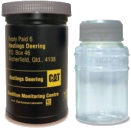
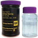
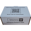
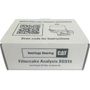
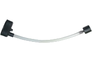
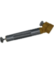
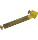
Understanding what goes on behind the scenes in your machine is critical to maximising machine reliability, productivity and reduced operating costs. Centrifugal oil filtration (COF) will potentially remove additional material that is traditionally detected by analysis of your oil. Hastings Deering have introduced an analysis to monitor the material captured in the filter cake of your COF systems. The SOS15 filter cake tool allows you to sample your filter cake in a consistent method for laboratory analysis.
Its simple all-in-one design allows for quick and easy sampling of your filter cake media. Microscopy analysis of the wear debris generated and captured in the filter cake can provide vital information of leading indicators for abnormal wear within your compartment. It is still recommended to continue with routine oil analysis in conjunction with this analysis.
For more information please contact Hastings Deering Laboratory Services.
Additional Testing
The laboratory also engages in project work from time to time where a customer's requirements do not fit in with the regular testing programs offered.
This would include the analysis of unusual or unidentified materials and consultancy services.